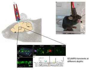
Our lab uses multiphoton microscopy to peer inside the brain for recording and modulating neural activity at the individual neuron level. We have developed a light-weight fiber-coupled head-mounted miniature two-photon microscope (< 4 grams) for imaging and stimulation in awake freely moving mice. Our miniature microscope employs electrowetting lenses for high-speed variable focusing with no moving parts in a compact design.
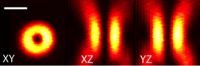
Superresolution microscopy
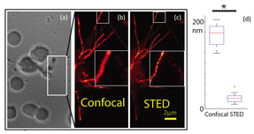
Our lab is developing Stimulated Emission Depletion (STED) microscopy for high-speed imaging of live neurons at resolutions of ~ 40-50 nm. In collaboration with Professor Diego Restrepo, we have used STED microscopy to image extremely small (< 100 nanometers) calcium microdomains in olfactory sensory neurons, important for signal transduction in olfaction. Additional neuroscience studies that can be performed using super resolution STED microscopy include studies of dendritic spines, involved in learning, myelin, important for problem solving, motor coordination and sensory input, synapses, key for transmission of the neural signal and primary cilia, important in neuronal differentiation.